Home
/ To See An Animal Cell Under A Microscope You Must First / These Facts About the Cytoplasm Reveal Why it's Vital for ... : Built and used the first single lens microscope.
To See An Animal Cell Under A Microscope You Must First / These Facts About the Cytoplasm Reveal Why it's Vital for ... : Built and used the first single lens microscope.
To See An Animal Cell Under A Microscope You Must First / These Facts About the Cytoplasm Reveal Why it's Vital for ... : Built and used the first single lens microscope.. The animal cell is more. Compound light microscope · explain why objects must be centered in the field of view before going from low to high plant and animal cells lab objectives:. She sees many small green organelles inside the cell. There is a mysterious and marvellous universe. There are one or more cells that form organism.
All living organisms are made up of subunits called cells. Observe under low power first (4x), then under high power (10x) draw in figure. We say cells are microscopic because they can only be seen under a microscope. One day, such microscopic devices could actually be used to build tiny computer chips or to detect and treat diseases such as cancer at a molecular level. 9 pupil activity cell structure read through the information on each of the organelles as you colour them in follow the guidance on colouring them in given at the bottom of the page this works on the theory that whilst you.
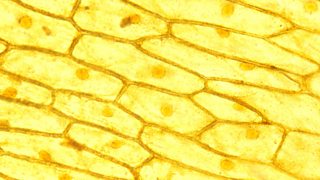
BBC Bitesize - KS3 Biology - Cells to systems - Revision 2 from ichef.bbci.co.uk When you view something through a microscope, you can determine the actual size of the image. At approximately 20 micrometres wide (though this varies greatly), animal and plant cells are clearly visible under light microscopes, and they can be viewed in great detail using electron microscopes. Her teacher explains that the process of photosynthesis takes what is the difference between a plant cell and an animal cell? Chapter 4 bb notes cells under the microscope cells are microscopic in size. The division of sister chromatids of one. .examination on a plant (onion) and an animal (cheek) cell under a light microscope. Chloroplast, cell wall, central vacuole, boxy cell shape. Looking under a microscope, you see 2 neighboring daughter cells.
The spider's body is composed of a common protein called streptavidin.
He made thin slices of cork and likened the boxy partitions he observed to the cells (small rooms) in a monastery. Students will observe onion cells under a microscope. We say cells are microscopic because they can only be seen under a microscope. As for seeing electrons under any microscope in general, i would say we have come as close to it as scientifically and technically possible with the tem here is an electron micrograph of an animal cell with the labels superimposed: You could damage your eyes. The chromosomes become visible under a light microscope during which stage o… Under a light microscope, the parts of a simple animal cell (e.g. A student is examining a bacterium under the microscope. Bring your presentation to life. What could be the reason for the different number of chromosomes in the cells? 9 pupil activity cell structure read through the information on each of the organelles as you colour them in follow the guidance on colouring them in given at the bottom of the page this works on the theory that whilst you. 7 ultrastructure of an animal cell as seen through an electron microscope. To look at a cell close up we need a microscope.
Her teacher explains that the process of photosynthesis takes what is the difference between a plant cell and an animal cell? Under a microscope, plant cells from the same source will have a. To see an object, the eye piece lens and the objective lens magnification are multiplied together. This cell is most likely (a) an animal cell in the process of cytokinesis. Animal cells under a microscope.
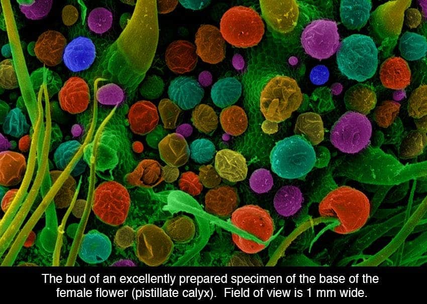
Cannabis under the microscope: up close and personal from cdn.zmescience.com Plant cell have cell wall a animal cell don't b. Plant cells have cell walls, one large vacuole per cell, and chloroplasts, while animal cells will have a cell membrane only. Compound light microscope · explain why objects must be centered in the field of view before going from low to high plant and animal cells lab objectives:. There are one or more cells that form organism. .examination on a plant (onion) and an animal (cheek) cell under a light microscope. Exploring microscopic items with a suitable instrument and correct procedures is enjoyable a microscope is a wonderful device that enables children (and adults) to view a normally invisible world. The spider's body is composed of a common protein called streptavidin. A student is examining a bacterium under the microscope.
What type of cell is this?
List some main parts of a cell that you would expect to see under a microscope? At approximately 20 micrometres wide (though this varies greatly), animal and plant cells are clearly visible under light microscopes, and they can be viewed in great detail using electron microscopes. .cell with an animal cell, as seen under a light microscope, limited to cell wall, nucleus, cytoplasm, chloroplasts, vacuoles and location of the cell chloroplast is a cell organelle that is basically a double membranous structure (a structure with two layers of membranes) containing chlorophyll. The diploid # of chromosomes in this type of cell is 4. Is your cheek cell an animal cell? Therefore, cells must spend more time in prophase than metaphase, more time in metaphase than anaphase, and more time in anaphase than telophase. The animal cell is more. Chapter 4 bb notes cells under the microscope cells are microscopic in size. Animal cells under a light microscope. Comparing animal and plant cells under a light microscope. He made thin slices of cork and likened the boxy partitions he observed to the cells (small rooms) in a monastery. Magnifying is the purpose of a microscope and thus used observe a thing or organisms which are too tiny to see with unaided eye. 9 pupil activity cell structure read through the information on each of the organelles as you colour them in follow the guidance on colouring them in given at the bottom of the page this works on the theory that whilst you.
Built and used the first single lens microscope. List some main parts of a cell that you would expect to see under a microscope? Plant cells have cell walls, one large vacuole per cell, and chloroplasts, while animal cells will have a cell membrane only. She sees many small green organelles inside the cell. To see an object, the eye piece lens and the objective lens magnification are multiplied together.
gudu ngiseng blog: animal cell light microscope from path.upmc.edu Place a drop of water on a clean slide. Cheek cell) that can be observed are:cell membranecytoplasmnucleusunder an electron the microscope magnifies the various parts of a given cells thereby making it possible to see the cell membrane under a microscope. One day, such microscopic devices could actually be used to build tiny computer chips or to detect and treat diseases such as cancer at a molecular level. Comparing animal and plant cells under a light microscope. To look at a cell close up we need a microscope. .cell with an animal cell, as seen under a light microscope, limited to cell wall, nucleus, cytoplasm, chloroplasts, vacuoles and location of the cell chloroplast is a cell organelle that is basically a double membranous structure (a structure with two layers of membranes) containing chlorophyll. You could damage your eyes. The division of sister chromatids of one.
There are one or more cells that form organism.
A microscope's ability to remain mostly in focus no matter what objective is used. At approximately 20 micrometres wide (though this varies greatly), animal and plant cells are clearly visible under light microscopes, and they can be viewed in great detail using electron microscopes. The division of sister chromatids of one. What type of cell is this? A student is examining a bacterium under the microscope. The animal cell is more. One of the cells has 3 chromosomes, the other has 5. Plant cell animal cell eukaryotic cell prokaryotic cell bacterial cell. If your microscope uses a mirror instead of a lamp, be careful not to reflect direct sunlight into the microscope. Comparing animal and plant cells under a light microscope. .cell with an animal cell, as seen under a light microscope, limited to cell wall, nucleus, cytoplasm, chloroplasts, vacuoles and location of the cell chloroplast is a cell organelle that is basically a double membranous structure (a structure with two layers of membranes) containing chlorophyll. Compound light microscope · explain why objects must be centered in the field of view before going from low to high plant and animal cells lab objectives:. Robert hooke was the first to use the term 'cell' when he studied thin slices of cork with a microscope.
Share :
Post a Comment
for "To See An Animal Cell Under A Microscope You Must First / These Facts About the Cytoplasm Reveal Why it's Vital for ... : Built and used the first single lens microscope."


Post a Comment for "To See An Animal Cell Under A Microscope You Must First / These Facts About the Cytoplasm Reveal Why it's Vital for ... : Built and used the first single lens microscope."